Have you ever looked closely at your eyes in a mirror — or seen a clear photo of yourself — and noticed a small brown spot on the colored part of your eye? You may have wondered whether it’s harmless or something that deserves attention.
These spots are often iris freckles: tiny, flat deposits of pigment that sit on the surface of the iris, the ring of color that makes your eyes blue, green, or brown. For the vast majority of people, they are completely benign. But understanding what they are, how they differ from other pigmented lesions, and when to have them evaluated is worth knowing.
What Are Iris Freckles?
The iris is the colored, ring-shaped tissue at the front of the eye. It controls the size of your pupil, regulating how much light enters. Like skin, the iris contains melanocytes — cells that produce melanin, the pigment responsible for color.
An iris freckle is a small, flat accumulation of melanin on the surface of the iris. Structurally, it is very thin and sits on top of the iris tissue rather than within it. Most are less than one millimeter across and do not distort or press on surrounding tissue.
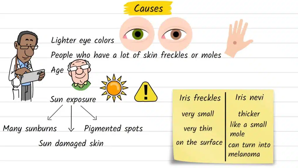
Iris freckles are distinct from iris nevi (the plural of nevus), which are deeper, thicker lesions — essentially moles within the iris tissue rather than on its surface. This anatomical difference matters clinically, which is covered further below.
Causes and Risk Factors
Iris freckles are not random. Research consistently points to a few key factors that make them more likely to develop.
Sun exposure is the most studied risk factor. A 2017 study published in Investigative Ophthalmology & Visual Science found that iris freckles were significantly more common in individuals with a history of sunburns, sun-damaged skin, and a high number of pigmented skin lesions. In that sense, they can serve as a subtle marker of cumulative ultraviolet light exposure over a person’s lifetime.
Other identified risk factors include:
- Light eye color — people with green, grey, or light brown eyes appear more susceptible
- Skin freckles and moles — a tendency toward melanin clustering in the skin seems to extend to the eyes
- Increasing age — iris freckles become more prevalent as people get older
- Genetic predisposition — a 2025 review in Investigative Ophthalmology & Visual Science identified genetic associations with iris freckling, suggesting hereditary components to pigment patterning in the eye
It’s worth noting that having iris freckles does not mean something is wrong. They are common findings in the general population, particularly among fair-skinned individuals.
Common Signs and Symptoms
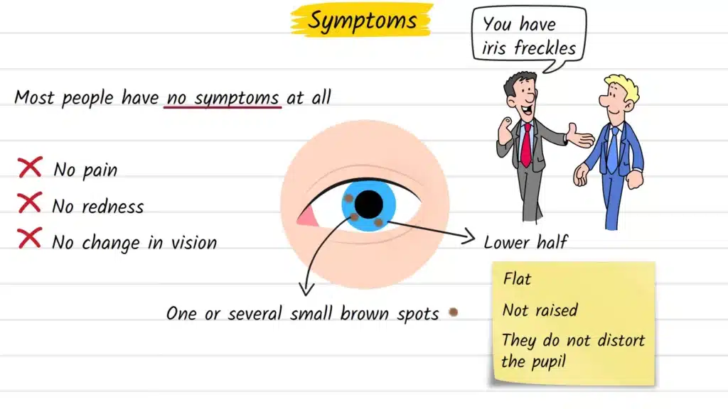
Most people with iris freckles experience no symptoms at all. There is typically no pain, redness, visual disturbance, or discomfort of any kind.
Many people discover them entirely by accident — during a routine eye exam, in a close-up photo, or when someone else notices them. What you might observe includes:
- One or more small, flat brown spots on the iris
- Spots that do not move, change color rapidly, or distort the pupil shape
- A tendency for freckles to cluster in the lower half of the iris, which receives more direct sun exposure than the upper portion (since the upper eyelid provides partial shade)
Because iris freckles cause no functional impairment, they are easy to overlook for years.
How the Condition Is Diagnosed
Diagnosing an iris freckle starts with a detailed examination at the slit lamp — the illuminated microscope your eye doctor uses during a standard eye exam. This instrument allows clinicians to view the iris in high magnification and assess:
- Size — freckles are typically under 1 mm in diameter
- Thickness — freckles are flat; they do not form a raised mass
- Location — freckles sit on the iris surface, not within the deeper stroma
- Tissue effect — they should not blur, compress, or distort the surrounding iris architecture
Distinguishing a freckle from an iris nevus requires careful observation. Nevi are larger, thicker, and may cause subtle architectural changes in nearby iris tissue. A 2024 paper in the British Journal of Ophthalmology emphasized that iris freckles deserve recognition as a clinically distinct entity — not simply a smaller version of a nevus.
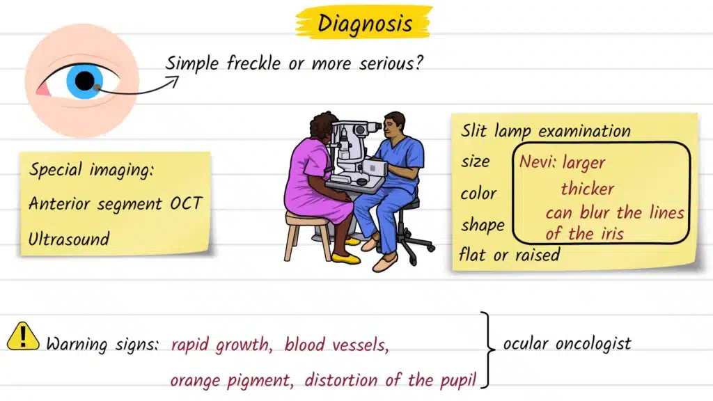
In cases where a lesion’s nature is unclear, additional imaging tools may be used:
- Anterior segment optical coherence tomography (AS-OCT) — a non-invasive scan that reveals the depth and layering of tissue
- Ultrasound biomicroscopy (UBM) — useful for assessing whether a lesion extends into deeper iris structures
If warning signs are present — such as rapid growth, the appearance of blood vessels on the lesion, orange pigment clumping (lipofuscin), or distortion of the pupil — a referral to an ocular oncologist for closer monitoring may be appropriate.
Treatment Options Explained
The straightforward answer: typical iris freckles require no treatment. They are monitored, not removed.
Management generally involves:
- Photographic documentation — your eye doctor may photograph the iris to establish a baseline and compare it at future visits
- Routine monitoring — annual or biennial eye exams to confirm the lesion remains stable
- Sun protection counseling — since UV exposure is a known contributing factor, patients are advised to wear quality sunglasses with UV400 or higher protection and wide-brimmed hats outdoors
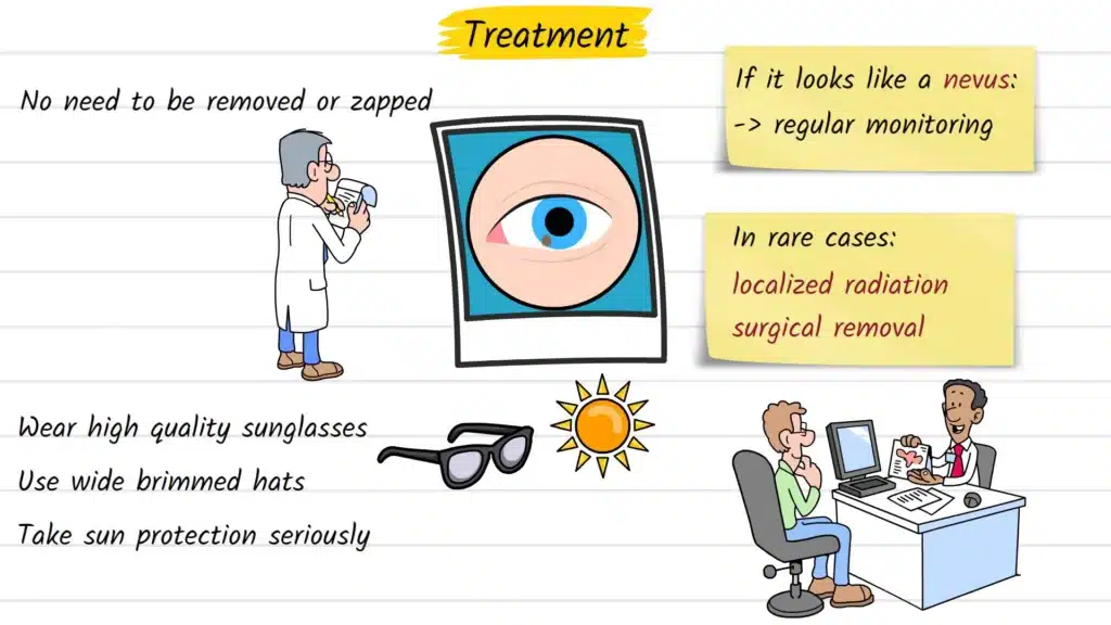
For iris nevi with higher-risk features, watchful waiting with structured follow-up is still the standard first approach. Intervention is reserved for rare cases where a lesion demonstrates growth or suspicious characteristics consistent with transformation. In those situations — which represent a small minority of cases — options may include localized plaque radiotherapy or, less commonly, surgical intervention. These decisions are made by specialist teams and depend on the individual clinical picture.
Possible Complications if Left Unmonitored
Iris freckles themselves carry no established risk of progression to cancer. They are surface lesions with no demonstrated malignant potential.
The clinical concern lies in accurate differentiation. Iris nevi — deeper lesions sometimes visually confused with freckles — do carry a very small risk of evolving into iris melanoma, a rare form of eye cancer. A 2003 study in the British Journal of Ophthalmology examined the association between posterior uveal melanoma and various iris pigmented lesions, underscoring the importance of correct classification.
Iris melanoma is rare, but when it does occur, early detection significantly improves outcomes. This is why eye doctors take pigmented iris lesions seriously — not to alarm patients, but to make sure nothing is missed.
The practical implication: if you have a pigmented spot on your iris that has never been formally evaluated, it is worth having it assessed once so it can be properly classified and documented.
Prevention and Risk Reduction
While you cannot change your genetics or eye color, there are evidence-backed steps to reduce UV-related stress on your eyes:
- Wear sunglasses labeled UV400 or with full UVA/UVB protection, especially during peak sun hours
- Choose wrap-around styles or lenses with large coverage for better peripheral protection
- Use a wide-brimmed hat as a complement to sunglasses — it reduces UV exposure reaching the eyes from above
- Avoid prolonged direct sun exposure during midday hours
- Apply this same protective thinking to your skin, since iris freckle risk correlates with broader UV-induced skin damage
These habits benefit overall eye health beyond iris pigmentation — reducing risk for cataracts, macular degeneration, and pterygium as well.
When to See an Eye Doctor
You should schedule an eye exam if:
- You notice a new or changing brown spot on your iris
- A spot appears to be growing in size or changing in color
- You develop any distortion in your field of vision, double vision, or sensitivity to light
- You have never had a formal eye exam to document an existing iris lesion
- You have a family or personal history of melanoma
Even without any of these concerns, a routine eye exam every one to two years is recommended for general ocular health. Many iris lesions are discovered incidentally — which is exactly when you want them found.
Frequently Asked Questions
In the vast majority of cases, no. Iris freckles are benign pigmented spots on the surface of the iris with no established risk of becoming cancerous. The key is distinguishing them from deeper lesions, such as iris nevi, which warrant monitoring.
An iris freckle is a thin, flat deposit of pigment sitting on the surface of the iris, typically under 1 mm. An iris nevus is deeper, thicker, and embedded within the iris tissue — more analogous to a mole in the skin. Eye doctors use slit lamp examination and sometimes imaging to tell them apart.
Iris freckles can appear or become more noticeable with age and cumulative sun exposure. However, rapid change in size, color, or shape is not expected and should be evaluated promptly by an eye care professional.
No. Iris freckles sit on the iris surface and do not interfere with the optical pathway. They do not affect visual acuity, contrast sensitivity, or any other aspect of vision.
Yes, though they are more commonly noted in adults. Children with light eye color or a genetic tendency toward freckling may develop them. Any pigmented iris lesion in a child should be reviewed by a pediatric ophthalmologist.
A personal or family history of melanoma is a reason to be more vigilant, not necessarily alarmed. Inform your eye doctor about your history so they can appropriately classify and monitor any iris lesions and tailor the follow-up schedule accordingly.
Iris melanoma is a rare malignant tumor arising from melanocytes within the iris. It typically presents as a larger, growing lesion — often with associated features like blood vessel formation, elevated intraocular pressure, or pupil distortion. It is in a completely different category from the flat, stable surface spots that define iris freckles.
Sources:
- Iris pigmented lesions: genetic basis of iris freckles and nevi. Invest Ophthalmol Vis Sci. 2025.
- Weis E, et al. Association between posterior uveal melanoma and iris freckles, iris naevi, and choroidal naevi. Br J Ophthalmol. 2003.
- Iris freckle — a distinct entity. Br J Ophthalmol. 2024.
- Kaliki S, et al. Iris melanoma: management and prognosis. Applied Sciences. 2020.
Medical Disclaimer
This article is intended for general educational purposes only. It does not constitute medical advice, diagnosis, or treatment. If you have concerns about a pigmented lesion on your eye, consult a qualified eye care professional. Only a licensed clinician can evaluate your individual situation.