Sign Up

Sign up to Eye Conditions Explained to ask questions, solve cases, and expand your knowledge. Create a profile and connect with eye-care professionals from all over the world. 🌎

Have an account? Sign In
Continue with Google
or use

Captcha Click on image to update the captcha.

Have an account? Sign In Now

Sign In

We want to empower everyone to share their knowledge for the benefit of the entire community. Let people who need your help find you.

Sign Up Here
Continue with Google
or use

Captcha Click on image to update the captcha.

Forgot Password?

Don't have account, Sign Up Here

Forgot Password

Lost your password? Please enter your email address and we will send a new Password at your email. You can change your Password any time from your profile.

Have an account? Sign In Now

Please briefly explain why you feel this question should be reported.

Please briefly explain why you feel this answer should be reported.

Please briefly explain why you feel this user should be reported.

Eye Conditions Explained Logo Eye Conditions Explained Logo
Sign InSign Up

Eye Conditions Explained

Eye Conditions Explained Navigation

Search
Ask A Question

Mobile menu

Close
  • Ask Questions
  • Read Articles
  • Explore Groups
  • View Categories
  • Login
What's your question?
  • Recent Questions
  • Most Answered
  • No Answers
  • Answers
  • Most Visited
  • Most Voted

Eye Conditions Explained Latest Questions

Vasilis Inglezis
  • 6
Poll
Vasilis Inglezis
Asked: February 16, 2019In: MCQs

What’s your diagnosis based on the image?

  • 6

Poll Results

8.42%Subhyaloid hemorrhage ( 47 voters )
12.37%Myelinated nerve fiber layer ( 69 voters )
67.56%Torpedo maculopathy ( 377 voters )
11.65%None of the above ( 65 voters )
Based On 558 Votes

Participate in Poll, Choose Your Answer.

What’s your diagnosis based on the image?

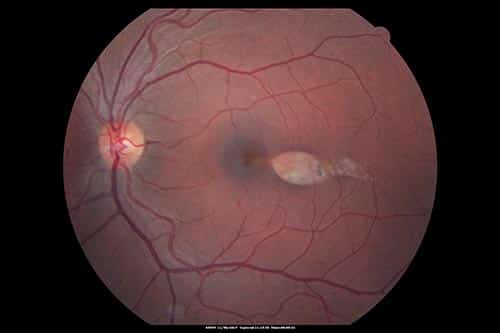
Similar question: How would you diagnose this based on the image?

myelinated nerve fiber layersubhyaloid hemorrhagetorpedo maculopathy
0
  • 0
  • 0
Vasilis Inglezis
  • 0
Poll
Vasilis Inglezis
Asked: January 31, 2019In: MCQs

The actions of inferior oblique muscle are:

  • 0

Poll Results

6.03%depression and adduction of the eye ( 34 voters )
14.36%intorsion, abduction and depression of the eye ( 81 voters )
18.09%elevation, adduction and intorsion of the eye ( 102 voters )
61.52%extorsion, elevation and abduction of the eye ( 347 voters )
Based On 564 Votes

Participate in Poll, Choose Your Answer.

Answer: Its actions are extorsion, elevation and abduction of the eye. Primary action is extorsion (external rotation); secondary action is ...Read more

extraocular muscles
0
  • 0
  • 0
Vasilis Inglezis
  • 1
Poll
Vasilis Inglezis
Asked: January 9, 2019In: MCQs

Which is the direction of the eye in oculomotor nerve palsy?

  • 1

Poll Results

7.8%It turns inwards ( 39 voters )
27%It turns outwards ( 135 voters )
18.2%It turns up and outwards ( 91 voters )
47%It turns down and outwards ( 235 voters )
Based On 500 Votes

Participate in Poll, Choose Your Answer.

2
  • 2
  • 0
Vasilis Inglezis
  • 0
Poll
Vasilis Inglezis
Asked: February 6, 2019In: MCQs

Perimetry commonly shows enlarged blind spots in case of:

  • 0

Poll Results

7.95%Usher syndrome ( 49 voters )
70.78%Papilledema ( 436 voters )
6.17%Vitreous hemorrhage ( 38 voters )
15.1%Retinal detachment ( 93 voters )
Based On 616 Votes

Participate in Poll, Choose Your Answer.

Related question: Which of the following statements is NOT true about cones:   Answer: Papilledema

blind spotpapilledemaperimetryretinal detachmentusher syndromevitreous hemorrhage
0
  • 0
  • 0
Vasilis Inglezis
  • 3
Poll
Vasilis Inglezis
Asked: January 7, 2019In: MCQs

Which test is contraindicated to do while testing an infant?

  • 3

Poll Results

65.89%The LogMAR chart ( 284 voters )
9.05%The Stycar language test ( 39 voters )
0.93%The Cardiff acuity test ( 4 voters )
14.15%The Sheridan Gardiner Test ( 61 voters )
Based On 431 Votes

Participate in Poll, Choose Your Answer.

1
  • 1
  • 0
Vasilis Inglezis
  • 0
Poll
Vasilis Inglezis
Asked: January 26, 2019In: MCQs

Which one of the following diagnostic tests is NOT useful in evaluating a patient with a retained magnetic intraocular foreign body?

  • 0

Poll Results

4.73%Ultrasonography ( 23 voters )
61.32%Magnetic resonance imaging (MRI) ( 298 voters )
17.28%Electrophysiology ( 84 voters )
16.67%Ophthalmoscopy ( 81 voters )
Based On 486 Votes

Participate in Poll, Choose Your Answer.

intraocular foreign bodymri
1
  • 1
  • 0
Vasilis Inglezis
  • 3
Poll
Vasilis Inglezis
Asked: January 27, 2019In: MCQs

Fleischer rings are indicative of:

  • 3

Poll Results

17.53%Fuchs' dystrophy ( 101 voters )
20.31%Iridocorneal endothelial syndrome ( 117 voters )
4.51%Eye injury ( 26 voters )
57.64%Keratoconus ( 332 voters )
Based On 576 Votes

Participate in Poll, Choose Your Answer.

fleischer rings
0
  • 0
  • 0

Sidebar

Stats

Recent Posts

  • Central Retinal Vein Occlusion: Symptoms & Treatment

  • Homonymous Hemianopia: Causes, Symptoms & Treatment

  • Iris Freckles: Causes, Diagnosis & When to See a Doctor

  • Papilledema: Optic Disc Swelling From High ICP

  • Iritis (Anterior Uveitis): Symptoms, Causes & Treatment

Top Groups

  • Eye Conditions MCQ Discussion Group
    • Public group

    Eye Conditions MCQ Discussion Group

Explore

  • Ask Questions
  • Read Articles
  • Explore Groups
  • View Categories
  • Login

© 2018-2026 Eye Conditions Explained. All Rights Reserved
Terms of Use.